Lateral lumbar spine radiograph with anterior wedge compression

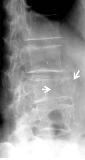
Download scientific diagram | Lateral lumbar spine radiograph with anterior wedge compression fractures of the L1, L2, and L4 vertebral bodies. from publication: Celiac Disease Presenting as Severe Osteopenia | The authors describe a unique presentation of celiac disease as multiple non-traumatic fractures in a young male without gastrointestinal complaints. A 29-year-old man presented with back pain and was found to have a non-traumatic compression fracture of the lumbar and | Celiac Disease, Osteopenia and Compression Fractures | ResearchGate, the professional network for scientists.
Typically Stable (Subsection 3A) - Clinical Imaging of Spinal Trauma
Spine SpringerLink
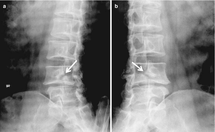
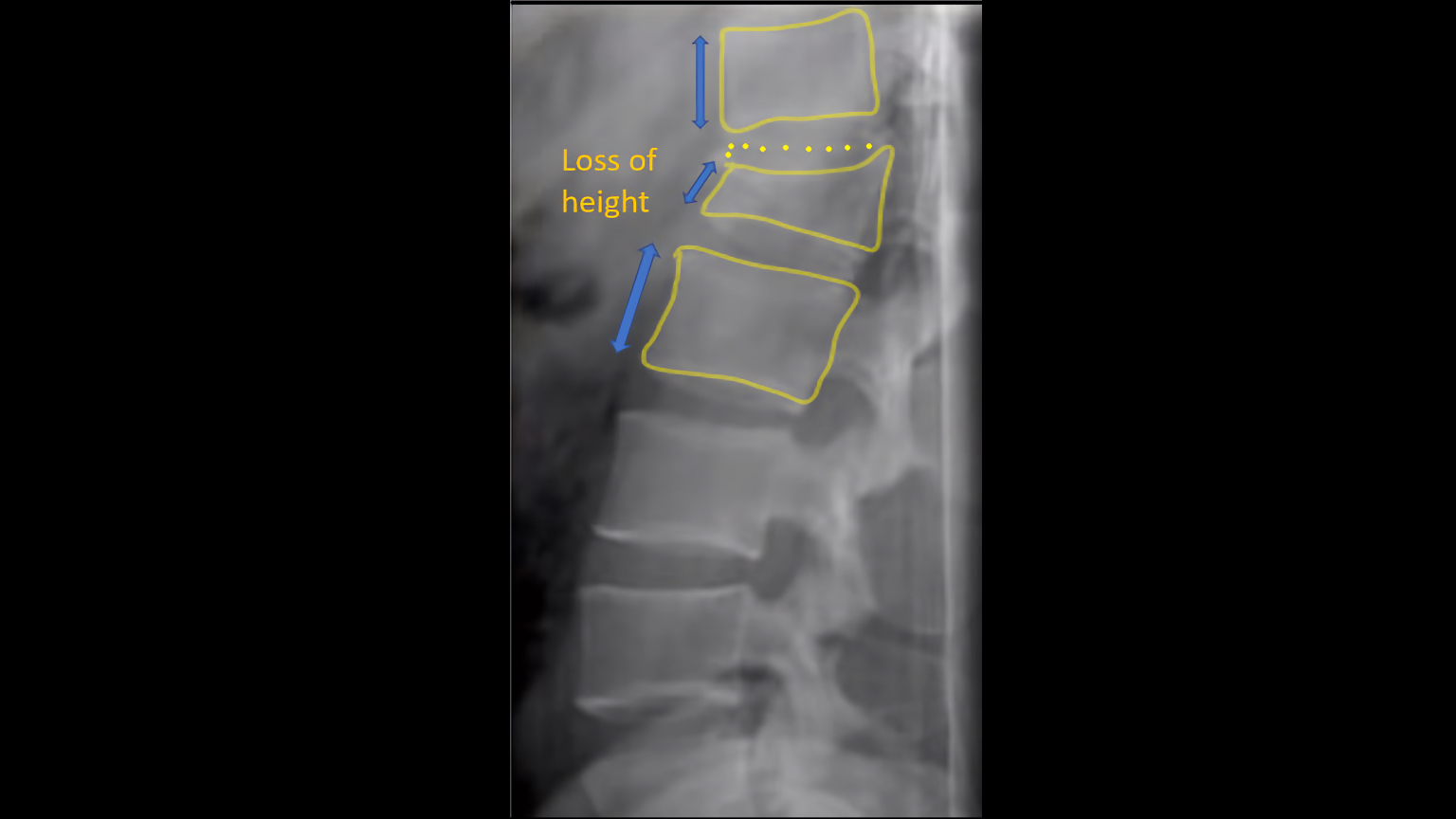
The correlation between vertebral wedge-shaped changes in X-ray imaging at supine and standing positions and the efficacy of operative treatment of thoracolumbar spinal fracture in the elderly

Vertebral fracture identification - ScienceDirect
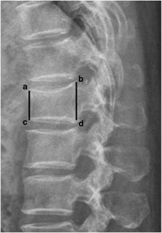
Lumbar X-ray Interpretation - OSCE Guide, Radiology
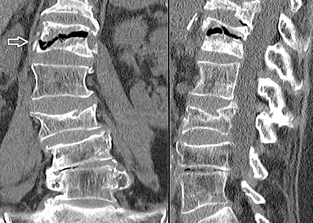
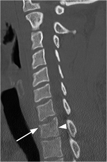
Identifying osteoporotic vertebral endplate and cortex fractures - Wáng - Quantitative Imaging in Medicine and Surgery

Diagnosis and Management of Vertebral Compression Fracture - ScienceDirect
Spine SpringerLink
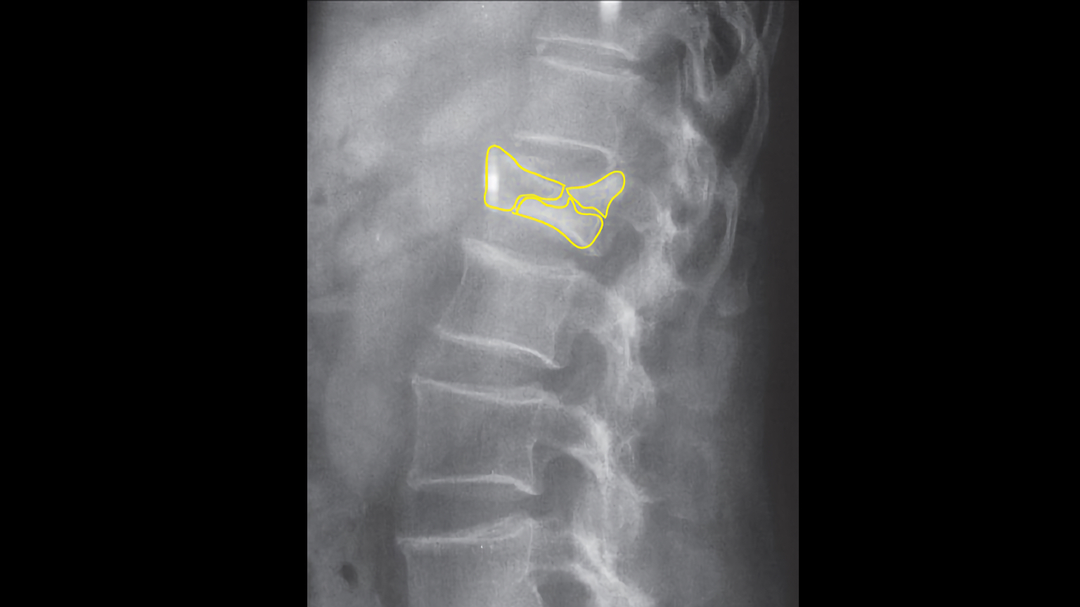
Difficult call. Distinguishing between physiological wedging of the

Radiology In Ped Emerg Med, Vol 5, Case 5
jcdr-12-BD03-g001.jpg

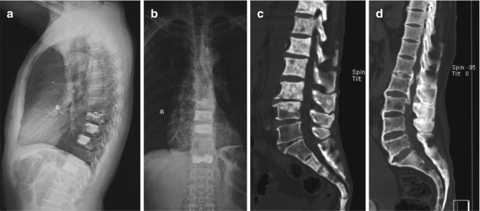
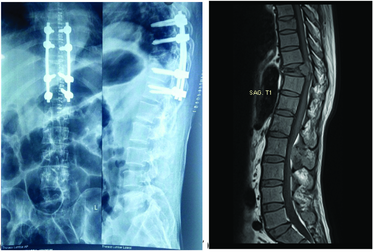
Case Study: T6 Vertebra Management in 58 Year Old Female
Spinal Injuries (Chapter 7) - Color Atlas of Emergency Trauma
Lumbar X-ray Interpretation - OSCE Guide, Radiology